
Background
AI is increasingly used in dermatology to support tasks ranging from the diagnosis of melanoma, basal cell carcinoma, and cutaneous squamous cell carcinoma to the characterization of chronic inflammatory skin diseases, helping clinicians interpret dermatological images more consistently and efficiently1,2.
Histopathology-confirmed imaging plays a crucial role in training and validating such systems, ensuring that model outputs are anchored in robust diagnostic ground truth.
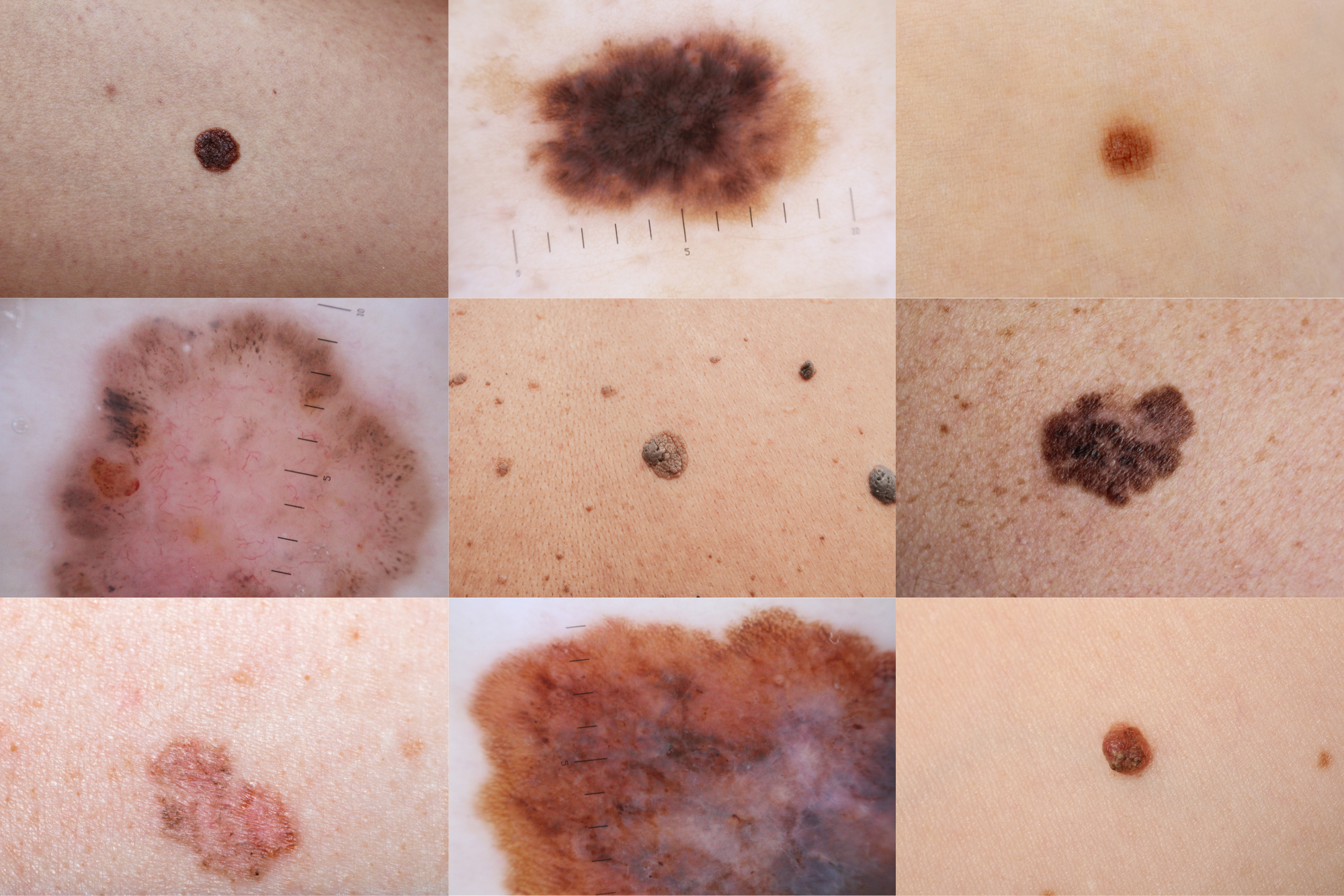
At the same time, real-world dermatology practice involves diverse image acquisition settings, making datasets that integrate macroscopic and dermoscopic images essential for clinically relevant AI.
Objective
A medical AI company engaged Aigora to curate a large, multimodal dermatology imaging dataset supporting the development and validation of AI algorithms for skin lesion detection and diagnosis.
Delivery
Aigora delivered a tailored multimodal imaging dataset combining clinical smartphone and dermoscopic images of the same lesions, linked to histopathologic diagnoses and structured metadata.
To learn more about Aigora’s experience in this field, read our research letter published in the Journal of the American Academy of Dermatology.